

Bronchogen 20mg
Bronchogen is a synthetic linear tetrapeptide composed of four amino acid residues arranged in the sequence Ala-Asp-Glu-Leu. Supplied as a lyophilized powder for laboratory research and analytical use only. Not intended for human or veterinary use, including consumption, injection, topical application, or any diagnostic, therapeutic, or clinical purpose.
Certificate of Analysis (COA)
Click “Check the COA” in the product section above to access the available Certificate of Analysis for this product. Please refer to the report for batch information and analytical results.
For laboratory research use only. Not for human or veterinary use.
Bronchogen Structure
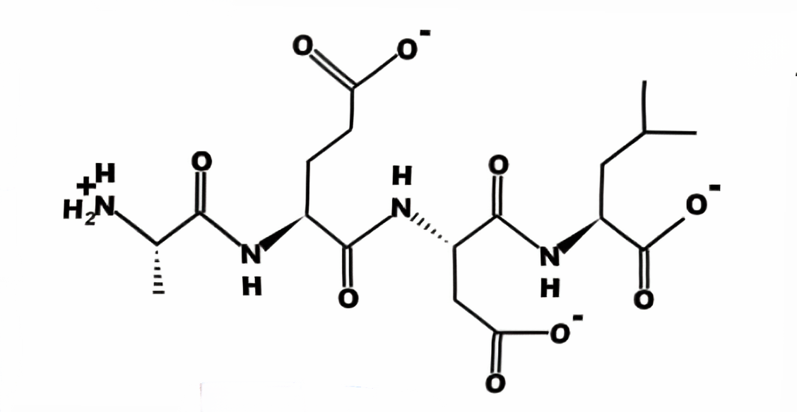 PubChem
- Sequence:Ala-Glu-Asp-Leu(AEDL)
- Molecular Formula:C18H30N4O9
- Molecular Weight:446.45g/mol
Bronchogen Research
Molecular Background
Bronchogen is a peptide discussed in nonclinical laboratory research; chemically speaking, it is a polypeptide with the amino acid sequence Ala-Asp-Glu-Leu—that is, a tetrapeptide[1].
Research Progress
J. R. Monaselidze estimated the thermodynamic parameters of DNA melting in the presence of Bronchogen at various concentrations using a differential scanning calorimeter. In this isolated-DNA experiment, the authors reported a stabilizing interaction with Bronchogen. The investigators measured an increase in the melting temperature of DNA from calf thymus and mouse liver by 3.1°C within a narrow range of r (molar ratio of bronchogen to DNA, 0.01–0.055). A further increase in r was not accompanied by changes in the melting temperature. The complex melting enthalpy (ΔHmelt) remained unchanged within this range of r (0.01–1.0). ΔHmelt for DNA from the thymus and mouse liver was 11.4 and 12.7 cal/g, respectively. The study authors interpreted these results as indicating that Bronchogen is not an adenine–thymine-specific or guanine–cytosine-specific ligand. The observed binding was characterized as strong and non-specific under the tested conditions. The authors also described binding to both strands of isolated DNA (primarily with nitrogen bases)[1].
Fig1. Dependence of DNA melting temperature on r at pH 7.02[1].
Major fraction (1); satellite fraction of calf thymus DNA (2). Major fraction (fine peak) of mouse liver DNA in solutions: (3) 0.015 M NaCl, 0.0015 M citrate buffer; (4) 0.15 M NaCl, 0.015 M citrate buffer.
In a nonclinical rat model of obstructive lung pathology, the effects of low-molecular-weight peptides on bronchial epithelial structure, barrier-related markers, and inflammatory measurements were investigated by N. A. Kuzubova. Chronic obstructive pulmonary disease was modeled in rats by 60-day intermittent exposure to NO2. In this rat model, one month of Bronchogen exposure was associated with changes in bronchial epithelial and lung tissue remodeling features (goblet cell hyperplasia, squamous metaplasia, lymphocytic infiltration and emphysema, and restoration of ciliated cells). The investigators also reported changes in secretory IgA, a local immunity marker, and bronchoalveolar cell composition and proinflammatory cytokine profiles consistent with lower measured neutrophilic inflammation in this rat model[3].
V. Kh. Khavinson measured the protein levels of Ki67, Mcl-1, p53, CD79, and NOS-3 in the 1st, 7th, and 14th passages of human embryonic bronchoepithelial cell cultures. Gene expression of NKX2-1, SCGB1A1, SCGB3A2, FOXA1, FOXA2, MUC4, MUC5AC, and SFTPA1 was measured by real-time polymerase chain reaction. Using spectrophotometry, viscometry, and circular dichroism, we studied the ADEL–DNA interaction in vitro. In these cell-culture experiments, the authors reported that ADEL was associated with changes in the levels of Ki67, Mcl-1, p53, CD79, and NOS-3 proteins in human bronchial epithelial cell cultures at various passages. The largest reported changes in Ki67- and Mcl-1-associated proliferation markers occurred in later-passage bronchial epithelial cell cultures. In this in vitro model, ADEL was reported to alter expression of genes involved in bronchial epithelial differentiation: NKX2-1, SCGB1A1, SCGB3A2, FOXA1, and FOXA2. The investigators also observed altered expression of genes discussed in relation to lung development: MUC4, MUC5AC, and SFTPA1. These laboratory methods were interpreted as evidence of ADEL–DNA interaction involving a region in the major groove (N7 guanine)[4].
Fig2. The influence of peptide ADEL on the gene expression of bronchial epithelium[4].
*p < 0.05—as compared to control
-
O.N. Titova investigated the effects of the tetrapeptide bronchogen on the structural and functional status of bronchial epithelium and pulmonary inflammatory activity in a rat model of chronic obstructive pulmonary disease (COPD) induced by 60 days of intermittent nitrogen dioxide exposure. The cell composition and cytokine-enzyme profile of bronchoalveolar lavage fluid (BALF), as well as the levels of secretory immunoglobulin A and surfactant protein B in BALF, were determined. During the experimental observation period, the authors reported lower measured neutrophilic inflammation and changes in bronchoalveolar cellular composition, cytokines, and enzymes in the treated rats. In this rat model, the investigators also described changes in previously disrupted bronchial epithelial structure and increases in secretory immunoglobulin A (a local immunity marker) and surfactant protein B (related to alveolar surface tension)[5].
Research by L. I. Fedoreyeva found that the exogenous short bioactive peptides epitalon (Ala-Glu-Asp-Gly), bronchogen (Ala-Glu-Asp-Leu), and vilon (Lys-Glu), at concentrations of 10⁻⁷–10⁻⁹ M, significantly affected the growth, development, and differentiation of the tobacco (Nicotiana tabacum) callus cultures. In these plant cultures, the researchers observed increased callus growth and leaf formation following exposure to epitalon and bronchogen. The study reported changes in gene expression in tobacco cells following peptide exposure, including genes responsible for tissue formation and cell differentiation. The authors also reported differential expression of CLE family genes—which encode known endogenous regulatory peptides—as well as the KNOX1 gene (a transcription factor gene) and the GRF gene (a growth regulatory factor gene), which encode corresponding DNA-binding proteins (such as topoisomerases and nucleases)[6].
Influence of Ala-Asp-Glu-Leu peptide on thermostability of white rat liver chromatin, in which H1 histone and non-histone proteins are depleted, have been studied by J Monaselidze. The authors reported that in 10 nm chromatin filaments, in which nucleosomes do not interact, peptide exposure was associated with altered nucleosomal core structure and release of approximately 15% of core DNA that melts in the melting temperature range of internucleosomal linker DNA. The authors suggested that these chromatin changes may increase DNA accessibility for transcription in the experimental system[2].
Research Limitations
It remains unclear whether the binding of Bronchogen to DNA is sequence-specific or structure-specific; in other words, it is not yet known whether Bronchogen binds randomly to the major or minor groove of DNA or whether it specifically recognizes certain promoter or enhancer regions. This characteristic of nonspecific binding makes it difficult to directly attribute the observed changes in gene expression (such as the upregulation of NKX2-1 and SCGB1A1) to specific DNA-peptide interactions[4]. These results derive from isolated DNA, cell cultures, plant cultures, and rat models; they do not establish safety or efficacy in humans. The plant-culture publication [6] describes Ala-Glu-Asp-Leu, which differs in residue order from the Ala-Asp-Glu-Leu sequence stated above; results for these sequences should not be assumed interchangeable.
COCER Peptides presents Bronchogen solely for nonclinical laboratory research. It is not for human or veterinary use, administration, or consumption by any route, and is not intended for diagnosis, treatment, prevention, or other clinical use. The above summary reports findings from cited nonclinical and in vitro studies only; it does not establish clinical safety, effectiveness, or FDA approval and provides no instructions for use.
Relevant Citations
References
[1] Monaselidze J R, Khavinson V K, Gorgoshidze M Z, et al. Effect of the Peptide Bronchogen (Ala-Asp-Glu-Leu) on DNA Thermostability[J]. Bulletin of Experimental Biology and Medicine, 2011,150(3):375-377.
DOI:10.1007/s10517-011-1146-x.
[2] Monaselidze J, Gorgoshidze M, Jokhadze T, et al. Influence of tetrapeptide on chromatin thermostability[J]. Georgian Med News, 2011(194):64-66.
PMID: 21685526
[3] Kuzubova N A, Lebedeva E S, Dvorakovskaya I V, et al. Modulating Effect of Peptide Therapy on the Morphofunctional State of Bronchial Epithelium in Rats with Obstructive Lung Pathology[J]. Bulletin of Experimental Biology and Medicine, 2015,159(5):685-688.
DOI:10.1007/s10517-015-3047-x.
[4] Khavinson V K, Tendler S M, Vanyushin B F, et al. Peptide Regulation of Gene Expression and Protein Synthesis in Bronchial Epithelium[J]. Lung, 2014,192(5):781-791.
DOI:10.1007/s00408-014-9620-7.
[5] Titova O N, Kuzubova N A, Lebedeva E S, et al. [ANTIINFLAMMATORY AND REGENERATIVE EFFECT OF PEPTIDE THERAPY IN THE MODEL OF OBSTRUCTIVE LUNG PATHOLOGY][J]. Ross Fiziol Zh Im I M Sechenova, 2017,103(2):201-208.
PMID: 30199201
[6] Fedoreyeva L I, Dilovarova T A, Ashapkin V V, et al. Short Exogenous Peptides Regulate Expression of CLE, KNOX1, and GRF Family Genes in Nicotiana tabacum[J]. Biochemistry (Mosc), 2017,82(4):521-528.
DOI:10.1134/S0006297917040149.
ALL PRODUCTS, ARTICLES, TECHNICAL MATERIALS, PRODUCT DESCRIPTIONS, AND OTHER INFORMATION PROVIDED ON THIS WEBSITE ARE INTENDED SOLELY FOR LEGITIMATE LABORATORY RESEARCH, INFORMATIONAL, AND EDUCATIONAL PURPOSES.
Products offered by Cocer Peptides are intended exclusively for in vitro and laboratory research and are NOT intended for human or veterinary use, human consumption, clinical use, diagnostic use, therapeutic use, compounding, self-administration, injection, or any other form of administration to humans or animals. These products are not intended to diagnose, treat, cure, mitigate, or prevent any disease or medical condition, nor are they intended to affect the structure or function of the human or animal body. Unless expressly stated otherwise, products offered on this website have not been evaluated or approved by the U.S. Food and Drug Administration (FDA) for human or veterinary use. Any references to published studies, biochemical pathways, molecular mechanisms, or experimental findings are provided solely for scientific discussion and research purposes and do not constitute medical advice, therapeutic claims, dosing guidance, or instructions for use. Purchasers are solely responsible for ensuring that all products are acquired, handled, stored, used, and disposed of in accordance with applicable laws, regulations, institutional requirements, and laboratory safety standards.